Schedule Anything
BayCare offers several convenient health care services that help get care you need to get back to feeling better.
You have a one in eight chance of developing breast cancer in your lifetime, and nearly 89 percent of women diagnosed do not have a family history of breast cancer. If discovered in its earliest stages, the chances of surviving breast cancer are good.
Until now, the best method of early detection has been digital mammography, using a specially designed camera and a computer to produce an image displayed on a high-resolution monitor. While digital mammography is still one of the most advanced technologies available, it is only a two-dimensional picture of the breast. Since the breast is composed of pockets of dense tissue surrounded by fat, the overlapping tissue in a digital image makes it difficult to see tiny spots called microcalcifications, and other subtle signs of early cancer.
To help diagnose breast cancer early, BayCare has added 3-D breast tomosynthesis to our breast health services.
Breast tomosynthesis allows radiologists to evaluate breast tissue one layer at a time. This exciting new technology has recently been FDA approved and is now available at most BayCare imaging locations that offer mammography.
Breast tomosynthesis may be used in conjunction with traditional digital mammography as part of your annual screening mammogram. Very low X-ray energy is used during the screening examination so your radiation experience is safely below the American College of Radiology (ACR) guidelines.
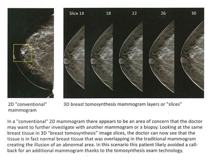
Breast tomosynthesis converts digital images into a stack of very thin layers or “slices,” building what is essentially a 3-D mammogram. This lets radiologists evaluate breast tissue one layer at a time, which allows them to detect 41 percent more invasive breast cancers and reduce false positives by up to 40 percent. This significantly reduces the number of times women are “called back” or asked to return for additional imaging.
During the tomosynthesis part of the exam, the X-ray arm sweeps in a slight arc over the breast, taking multiple breast images in just seconds. A computer then produces a 3-D image of your breast tissue in one millimeter layers. Radiologists can now see breast tissue in a more detailed way. Instead of viewing your breast tissue in a flat image, the tissue can be examined a millimeter at a time and fine details are more clearly visible.
A 3-D tomosynthesis exam is very similar to, and takes approximately as long as, a traditional mammogram. Just as with a digital mammogram, the technologist will position you, compress your breast under a paddle and take images from different angles. During the tomosynthesis portion of the exam, your breast will be under compression while the X-ray arm of the mammography machine makes a quick arc over the breast, taking a series of images from a number of angles. This takes only a few seconds and all of the images are viewed by the technologist to ensure they have captured adequate images for review by a radiologist.
The whole procedure takes approximately the same amount of time as that of a digital mammogram alone. The technologist sends your images electronically to the radiologist, who studies them and reports the results to your physician.
Make the clear choice when it comes to your breast health. Choose BayCare.