All of the BayCarePlus plans bundle your hospital, medical, and prescription drug benefits into one plan.
404 Error. Page Not Found
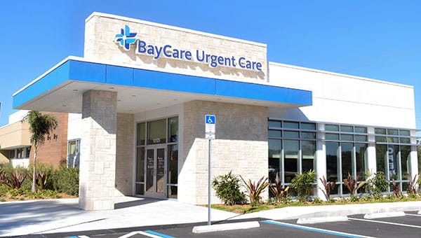
BayCare Urgent Care Centers are located throughout Tampa Bay and the surrounding areas. Find the location closest to you and Save Your Spot® online.
There are dozens of BayCare Lab locations throughout Tampa Bay and the surrounding areas. Find the BayCare Lab closest to you. You can also get directions, see lab hours, and Save Your Spot® online.
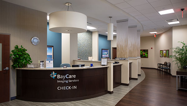
BayCare has several imaging centers located in and around Tampa Bay. Find an imaging center that is convenient for you and schedule your exam online.